
Image contest 2019 – The winners!
Light microscopy
Svetlana Pasteuning-Vuhman
Title: Neuronal sunflowers
Microscope: Confocal microscope LSM 880.
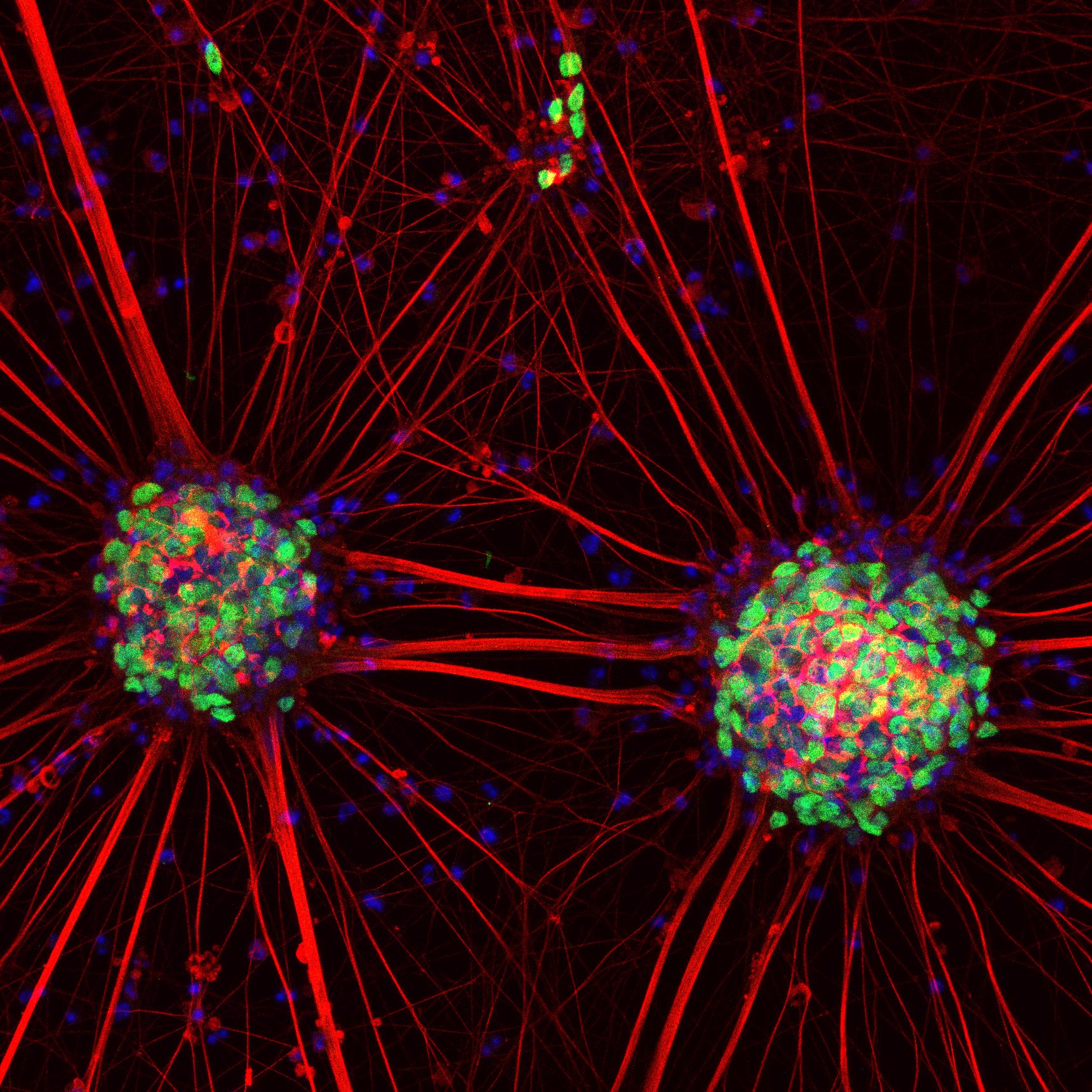
Description: Motor neurons were derived from human induced pluripotent stem cells and stained with neuronal markers.
Lotte Herstel
Title: Spiral of inhibitory neurons
Microscope: Confocal Zeiss LSM700
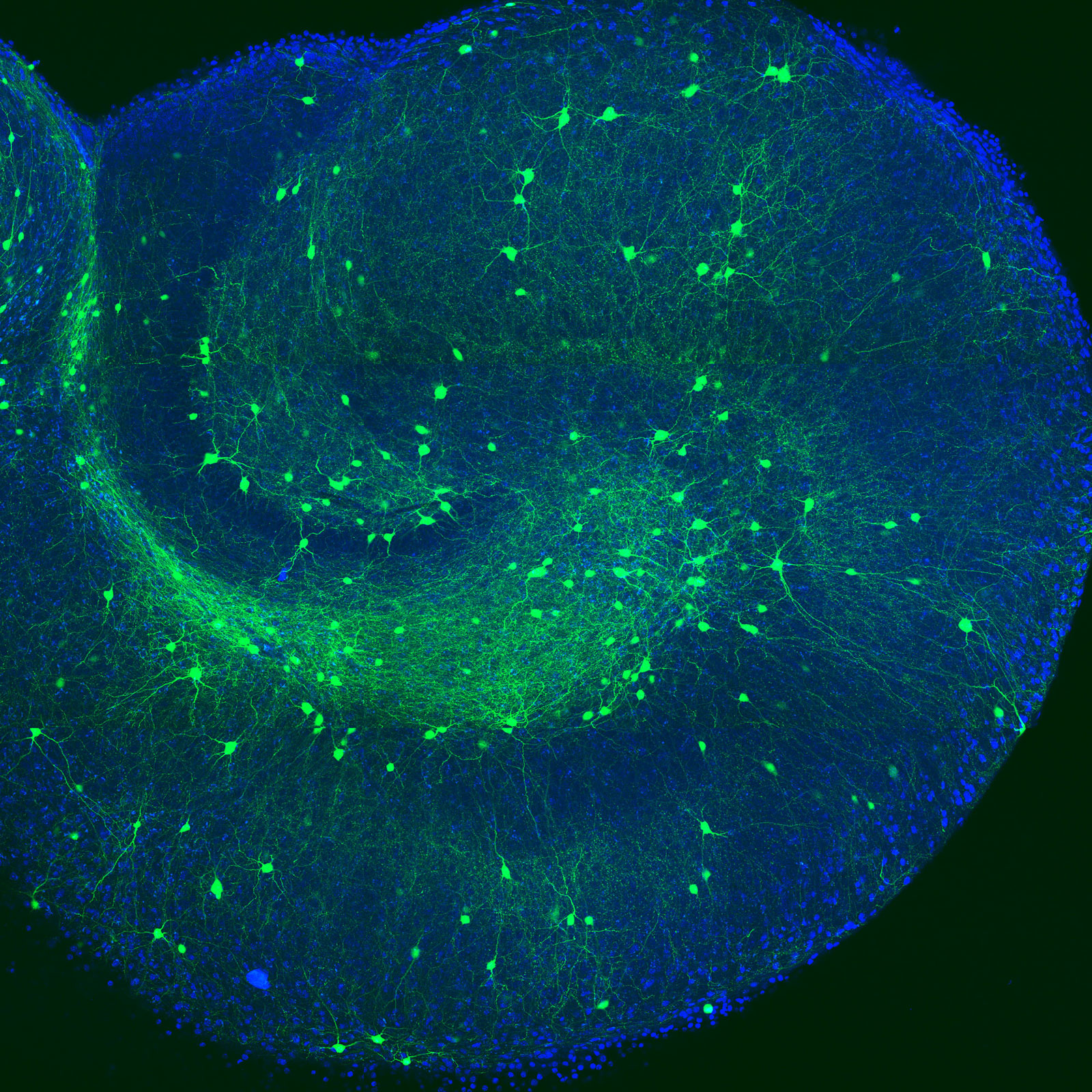
Description: Here shown is an organotypic hippocampal slice of a mouse brain, which was kept in culture for over 2 weeks before fixation. Labelled in blue are all cell bodies (DAPI), to show the outer contours of the slice. In green is the genetic labelling of GAD65-GFP, indicating a subset of inhibitory neurons that express this protein.
Wilco Nijenhuis
Title: Actin everywhere!
Microscope: Leica TCS SP8 STED 3X, 93x/1.30 glycerol objective
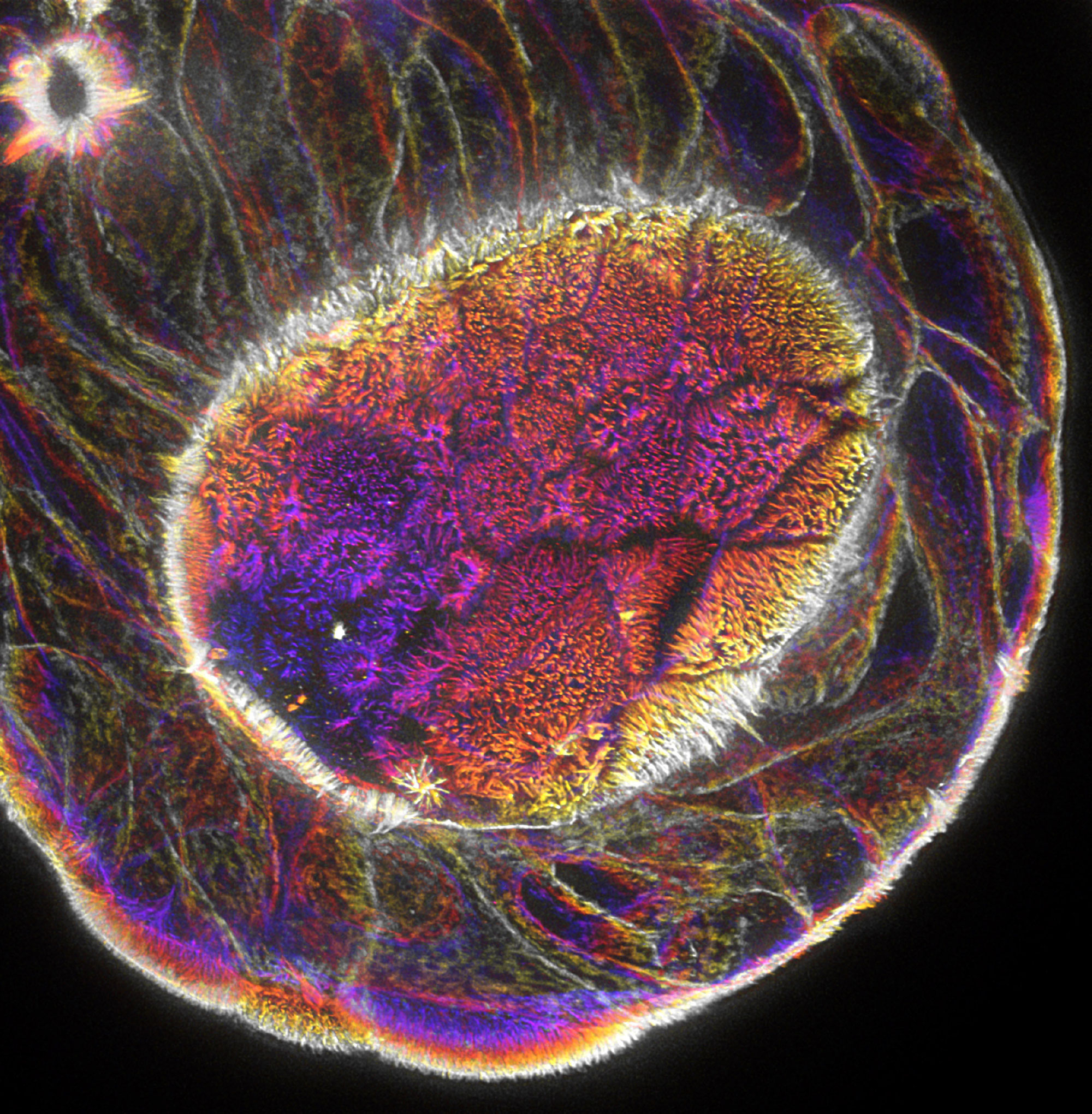
Description: STED image of actin in an human intestinal organoid resolving individual microvilli, depth encoded Z projection.
Babet van der Vaart
Title: My heart beats for science
Microscope: Spinning Disc 1 Nikon-Roper ILAS PhotoAblation (Kruyt building)
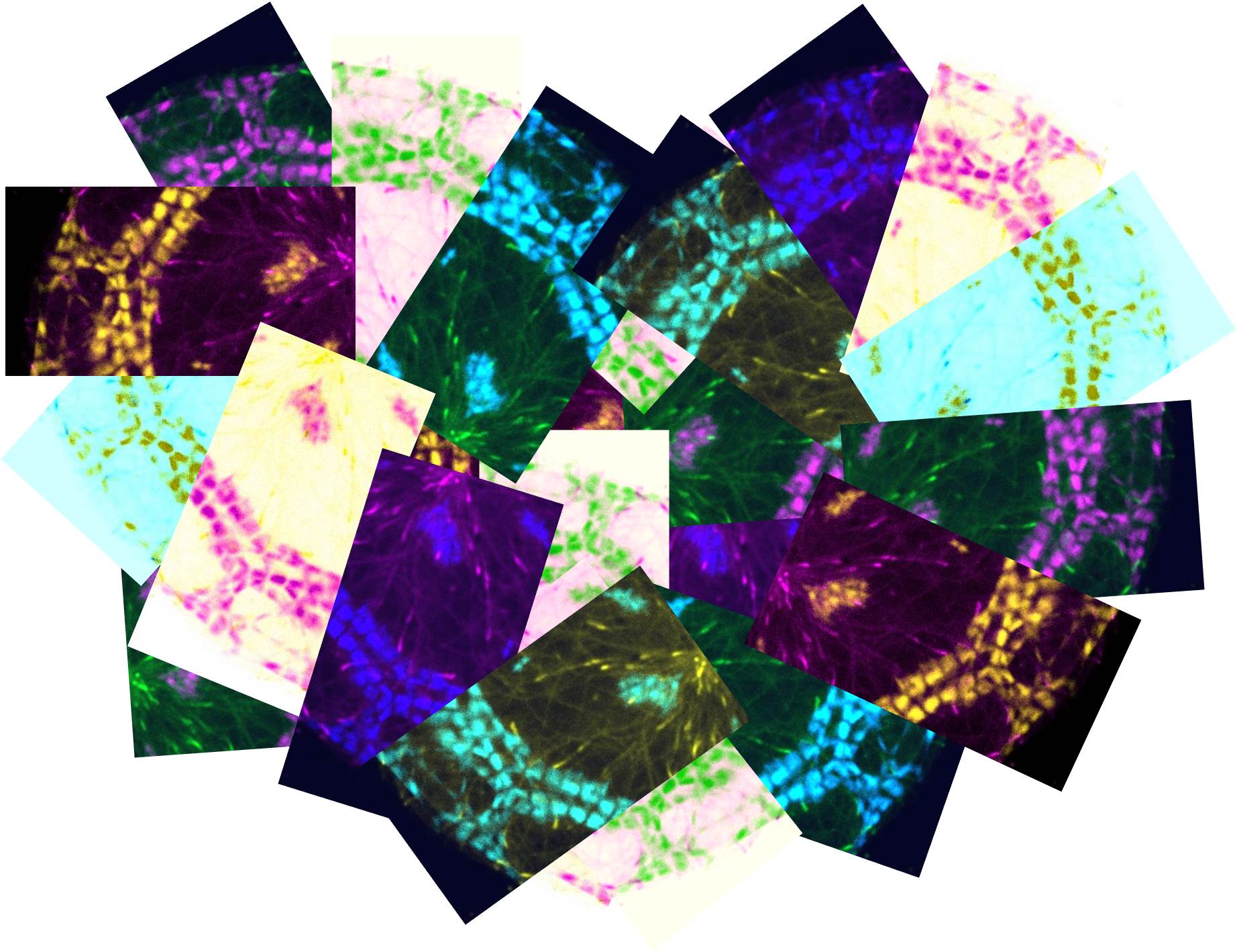
Description: This is a compilation of pictures to form a heart shape. The pictures show a zoom of a human induced-pluripotent stem cell-derived cardiomyocyte, which was transduced with lentiviral vectors to express a GFP-tagged microtubule plus-end marker (end-binding protein 3 (EB3)) and tagRFP-LifeAct; an actin marker. In cardiac cells the actin cytoskeleton forms contractile units named sarcomeres, which allow the cell to beat.
Electron microscopy
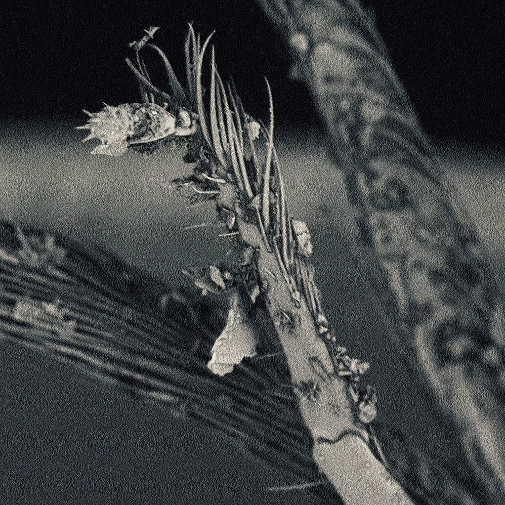
Xudong Ouyang
Title: Magic Forest
Microscope: Phenom PRO desktop scanning electron microscope
Description: It is a 360x magnification of antennas of a bee
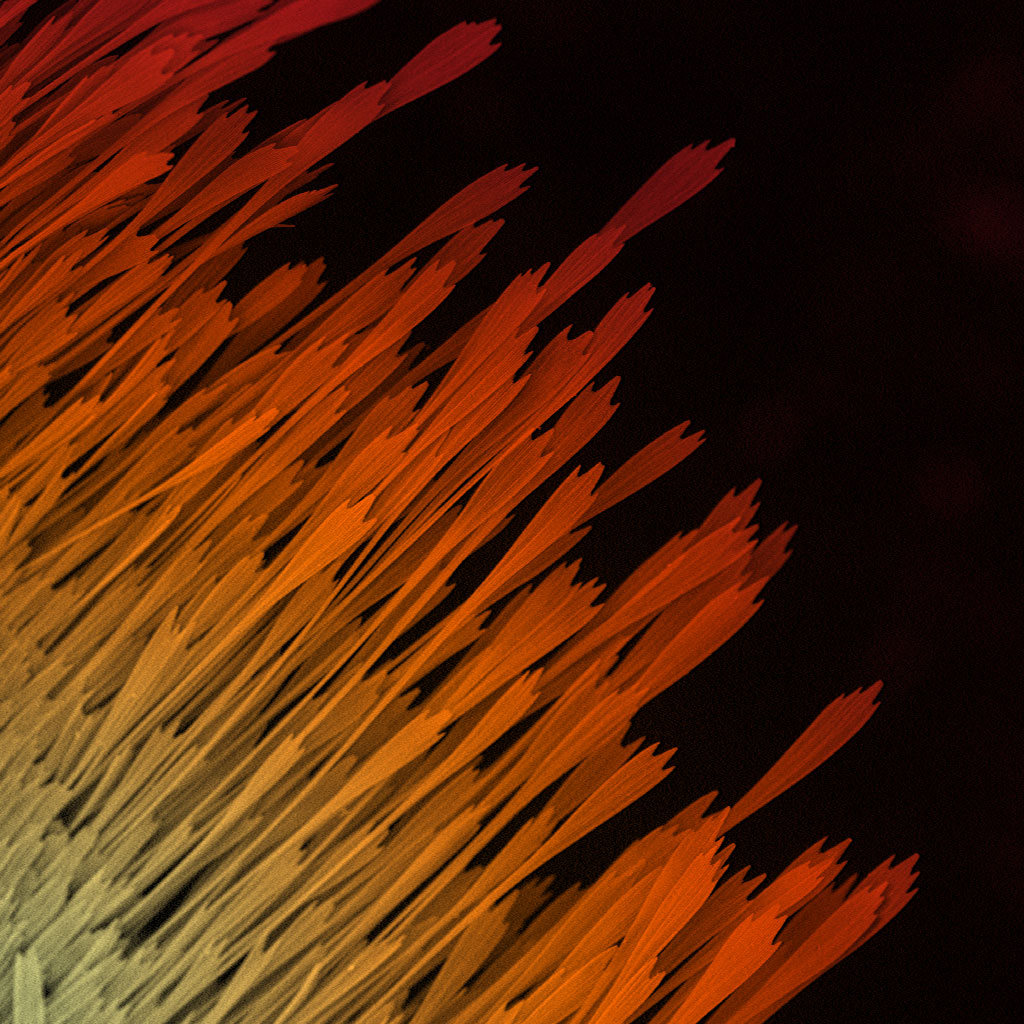
Samantha van der Beek
Title: Rising Phoenix
Microscope: Phenom PRO desktop scanning electron microscope
Description: A 320x magnification of the wing of a moth

Ann De Mazière
Title: Air waves
Microscope: JEOL 1011 electron microscope
Description: EM image of a part of a tracheole, a branch of the insect airway system, between the circular and longitudinal muscle cell layers of the Drosophila intestinal wall