Image contest 2015 – The winners!
Light microscopy
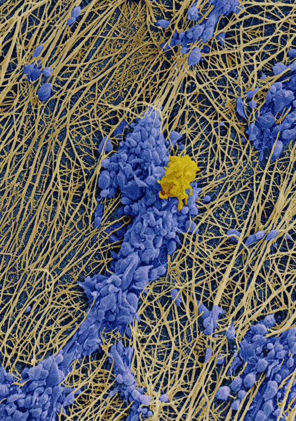
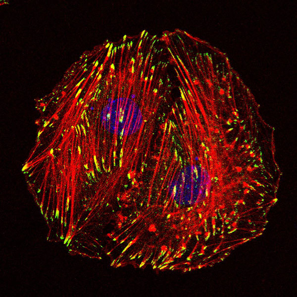 Chao Yang (Cell Biology, Department of Biology, Faculty of Science, Utrecht University)
The artistic name of the image: LOOK
Description: what the actin and focal adhesion looks like in A7R5 cells.
Microscope: Upright FL microscope Nikon Eclipse Ni
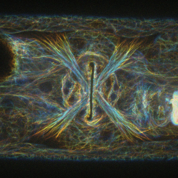 Mike Boxem (Developmental Biology, Department of Biology, Faculty of Science, Utrecht University)
The artistic name of the image: ‘evil eye’.
Description: It is an image of the C. elegans vulva (ventral view), in a line expressing endogenously (CRISPR/Cas9) tagged GFP::MAPH1. MAPH-1 is a C. elegans homolog of mammalian MAP1 microtuble associated proteins. The image is a color coded Z-stack of 10 slices (0.5 um apart).
Microscope: Andor/Nikon spinning disc confocal.
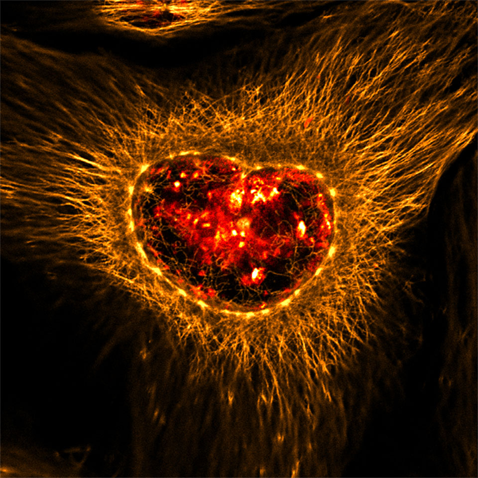
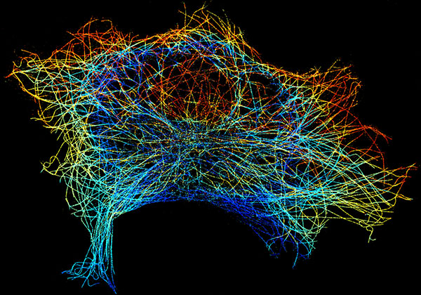 Bas Cloin (Cell Biology, Department of Biology, Faculty of Science, Utrecht University)
The artistic name of the image: Superduper microtubule network
Description: 3D superresolution reconstruction of the microtubule network of a complete COS7-cell made using DNA-paint and adaptive optics. Z-position is color-coded from red at the bottom to blue at the top.
Microscope: Nanoscope II
Electron microscopy
 René Scriwanek (UMC Utrecht) Clotting: Scanning electron micrograph of human blood platelets adhering to subendothelium under flow conditions
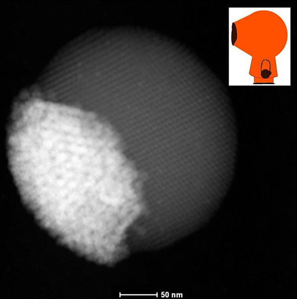 Da Wang (Soft Condensed Matter, Debye Institute for Nanomaterials Science, Utrecht University)
The artistic name of the image: Kenny McCormick’s head—Janus Binary supraparticle
Description: Self-assembled “Janus Binary Supraparticle” composed of Ag nanocrystals and Fe3O4 nanocrystals. Highlight: Janus binary supraparticle is first time found by our group. HAADF-STEM image of Janus binary supraparticle composed of Ag nanocrystals (bright part) and Fe3O4 nanocrystals (grey part) was acquired from Tecnai 20F microscope in Utrecht University. Fe3O4 rich part was highly ordered. The Janus binary supraparticle looked like the side view of Kenny McCormick’s head (Inset picture, see cartoon “South Park”).
Microscope: Tecnai 20F (FEI company), in HAADF-STEM mode
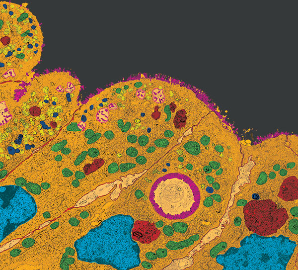 Kerstin Schneeberger (UMC Utrecht)
The artistic name of the image: The Introverted Intestine
Description: The image shows intestinal enterocytes of a mouse model for microvillus inclusion disease. Upon deletion of Myo5b, the mice develop severe diarrhea, which is caused by defective trafficking and polarity in the enterocytes. Instead of having microvilli and transporters on the apical membrane, diseased enterocytes display an accumulation of apical proteins, endosomes and microvillus inclusions in the cytoplasma.
Microscope: Jeol 1010 electron microscope (UMC Utrecht)
These images won the prize last year
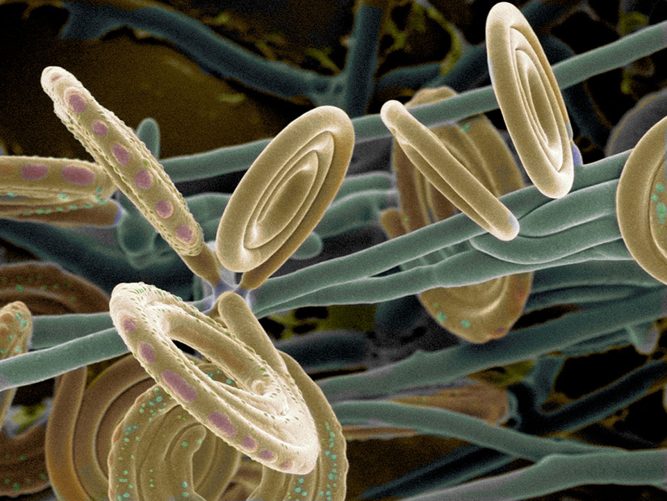
Will you be the next winner?
For the fourth year the Bio-Imaging Utrecht workgroup organizes a microscope image competition. The focus of this competition is to show that microscopic images often not only have scientific value but also an artistic value, the images will be judged on their visual effect.
Any microscope image can participate in the competition. We have two categories: Fluorescence images and Electron microscopy images. The first price will be 200 euro.
A few rules apply:
- One image per person can be submitted.
- Mild processing is allowed.
- The image must be made with equipment present in Utrecht.
Image format: JPEG
Deadline: October 15th, 2015
How to participate: send your image together with an artistic title, name of the machine that was used and a short scientific description to: meetings@bioimaging-utrecht.nl
Utrecht Microscopy Image Contest 2015
Livio Kleij (Molecular Cancer Research, UMCU)
Anko de Graaff (Hubrecht imaging centre, Hubrecht)
Ilya Grigoriev ( Cell Biology, UU)
Corlinda ten Brink (Cell Biology, UMCU)
Dave van den Heuvel (Molecular Biophysics, UU)