2019 Image Contest
Light microscopy
LM01
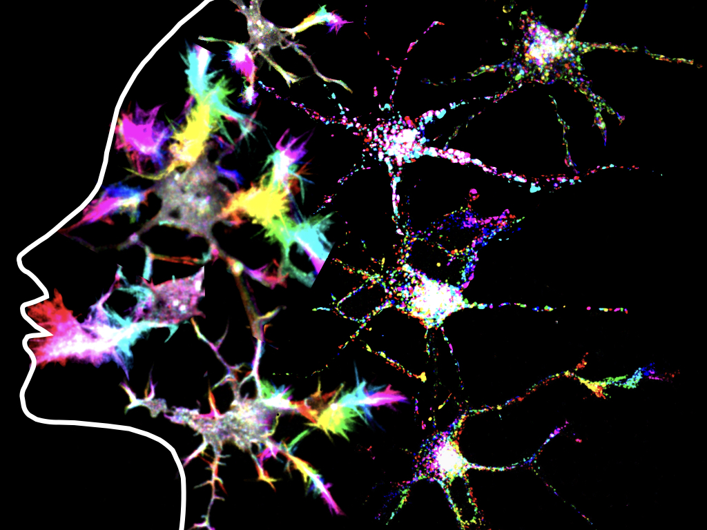
Neuron psychedelia
microscope: Nikon spinning disk
description: Time color-coded dynamics of actin and lysosomes in young hippocampal neurons
Author: Mithila Burute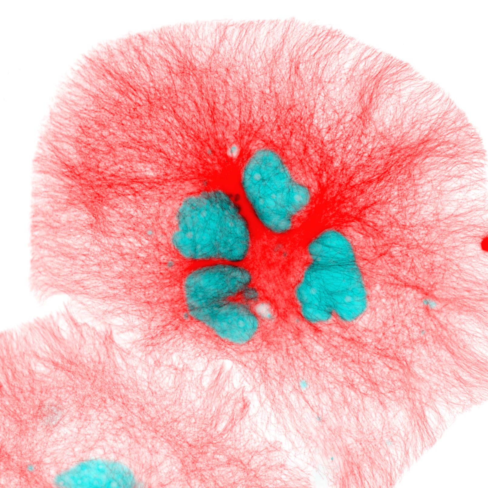
LM02
Spirit and Tranquil
microscope: Nikon Upright Microscope
description: The human retinal pigment epithelium (hRPE) cells that treated with Centrinone, a compound that inhibit the mature of centriole, lost the ability to form microtubule organization center, thus leads to a failure to form the spindle and to dividing; and the cells changed to multi-nuclear cells. Red: microtubule, Cyan: nuclear
Author: Fangrui Chen
LM03
Hubrecht
microscope: Confocal Leica SP8
description: Whole-mount staining of a pancreatic organoid. The organoid is shaped like an embryo (Hubrecht logo). Therefore I will simply call it “Hubrecht”
Author: Tim Dielen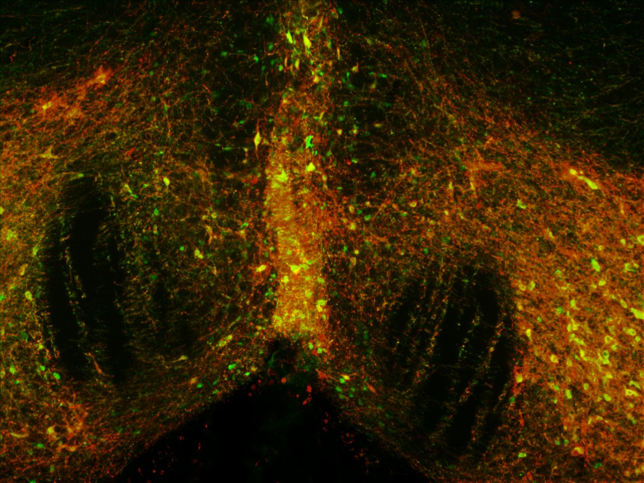
LM04
Phoenix
microscope: Olympus BX60 Fluorescence microscope
description: TH and DsRed double staining in brain slices of rats treated with DREADD technology
Author: Judith Hendriks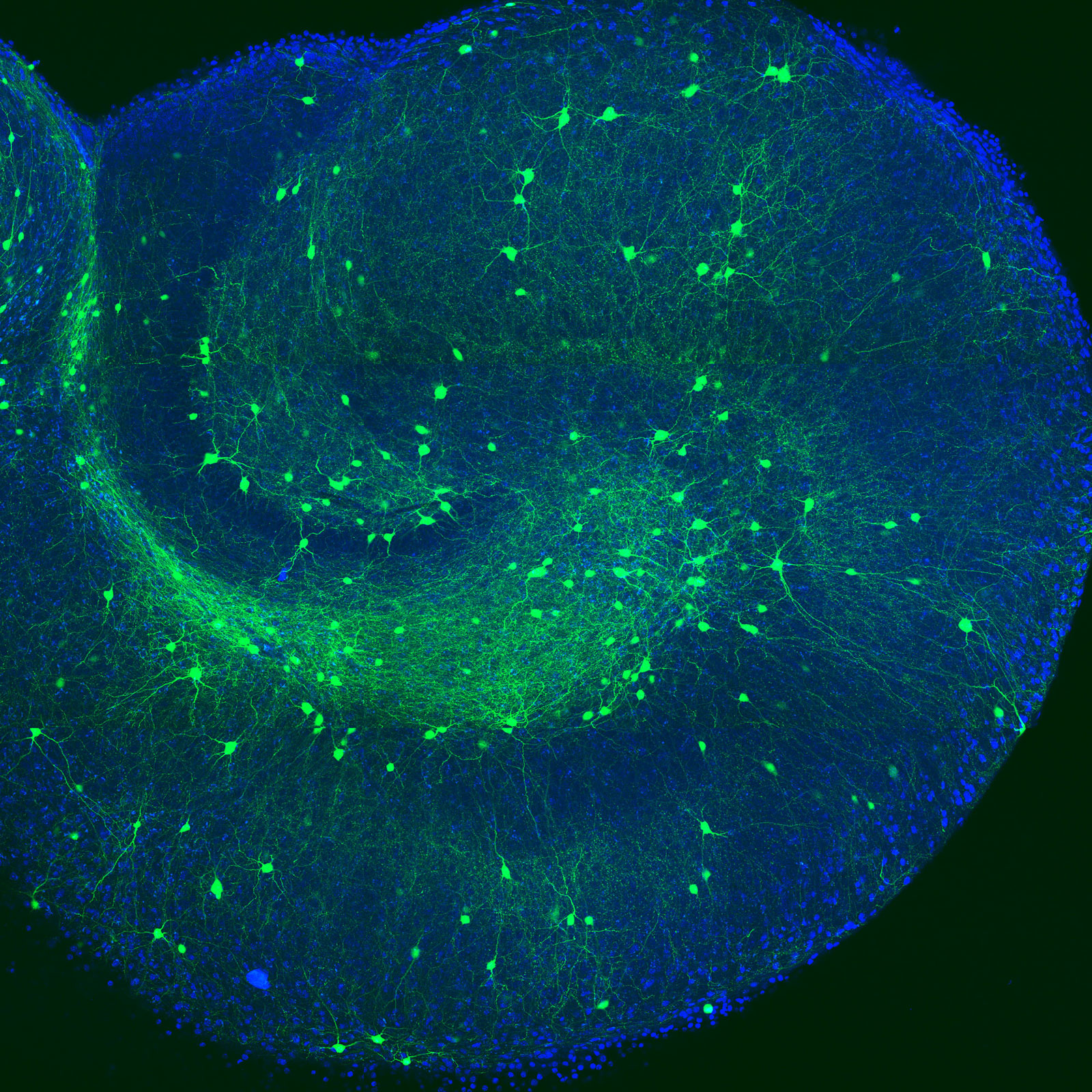
LM05
Spiral of inhibitory neurons
microscope: Confocal Zeiss LSM700
description: Here shown is an organotypic hippocampal slice of a mouse brain, which was kept in culture for over 2 weeks before fixation. Labelled in blue are all cell bodies (DAPI), to show the outer contours of the slice. In green is the genetic labelling of GAD65-GFP, indicating a subset of inhibitory neurons that express this protein
Author: Lotte Herstel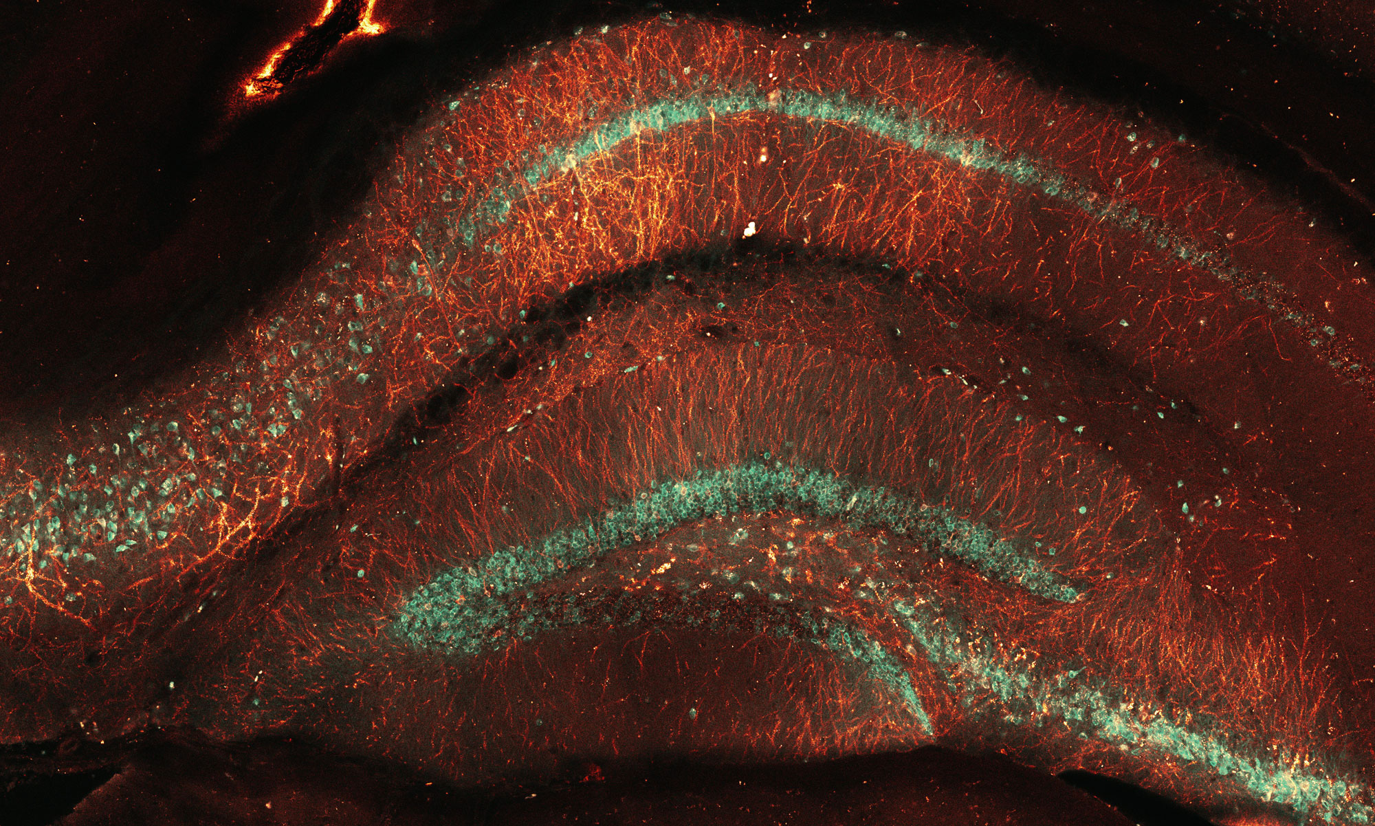
LM06
Knockin’ in memories
microscope: Zeiss LSM700
description: CRISPR knock-in for PSD95, in the hippocampal formation of mouse brain
Author: Arthur Jong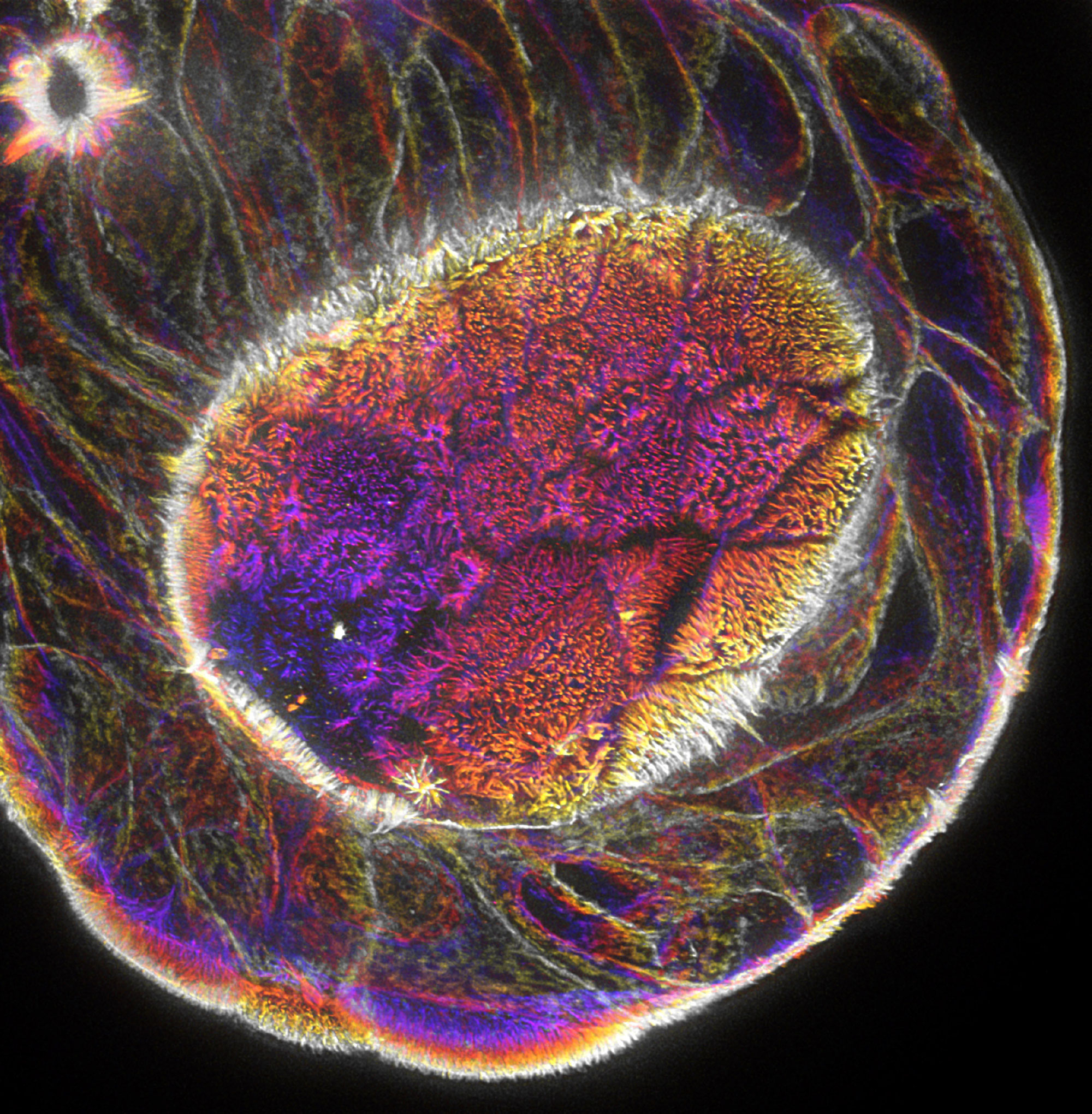
LM07
Actin everywhere!
microscope: Leica TCS SP8 STED 3X, 93x/1.30 glycerol objective
description: STED image of actin in an human intestinal organoid resolving individual microvilli, depth encoded Z projection
Author: Wilco Nijenhuis
LM08
Night of the globular waves
microscope: Leica DMI 6000 CS AFC (STED)
description: Spreading of breast cancer cells on a 2D surface. The image shows the cytoskeletal fibers of the cells which are polymers of globular proteins. Microtubules (red), F-actin (cyan), nuclei (blue).
Author: Milena Pasolli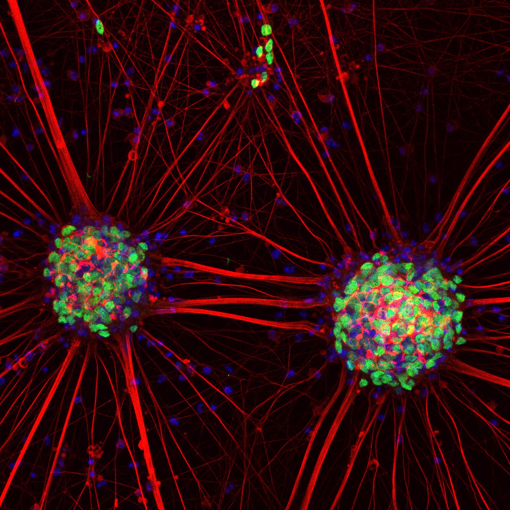
LM09
Neuronal sunflowers
microscope: Confocal microscope LSM 880.
description: Motor neurons were derived from human induced pluripotent stem cells and stained with neuronal markers
Author: Svetlana Pasteuning-Vuhman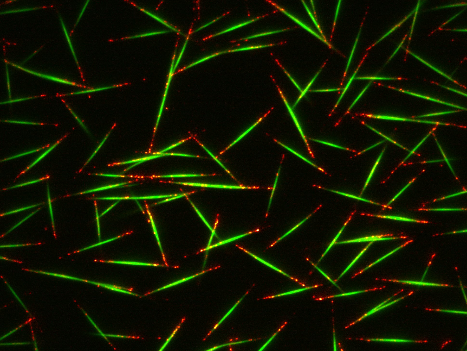
LM10
Shooting Stars
microscope: TIRF Nikon Ti – Roper iLas-2 FRAP/Photoablation system
description: Image shows single frame of anti-parallel microtubule bundles reconstituted in vitro on a glass surface. The idea of this experiment was to create bundles of two anti-parallel microtubules by adding a microtubule bundling protein PRC1 (green). However, a little bit higher concentration of PRC1 produced bundles of multiple microtubules of similar size (depicted by red comets, marked by microtubule plus-end marker EB3)
Author: Dipti Rai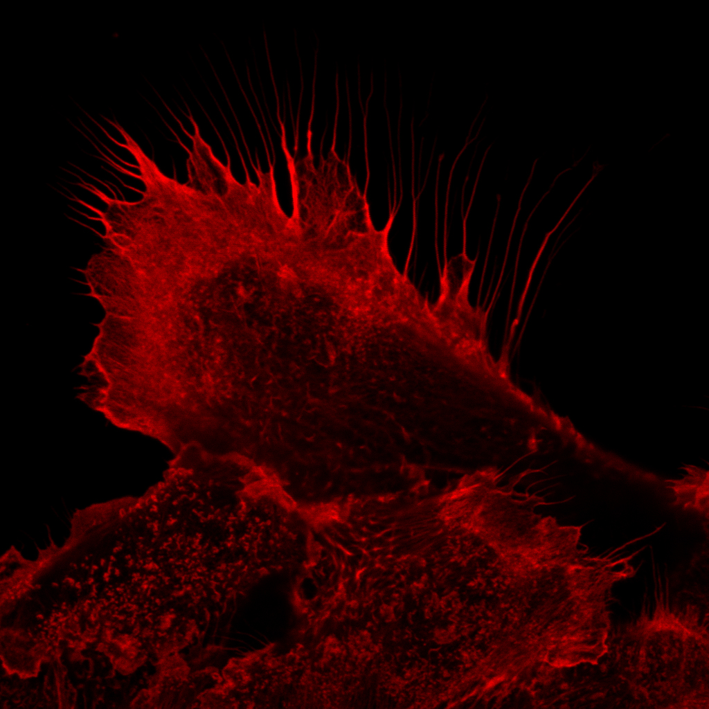
LM11
Hairy Pancreatic Cancer Cell
microscope: ZEISS LSM700
description: F-actin staining of strongly adherent Pancreatic Ductal Adenocarcinoma cells
Author: Joep Sprangers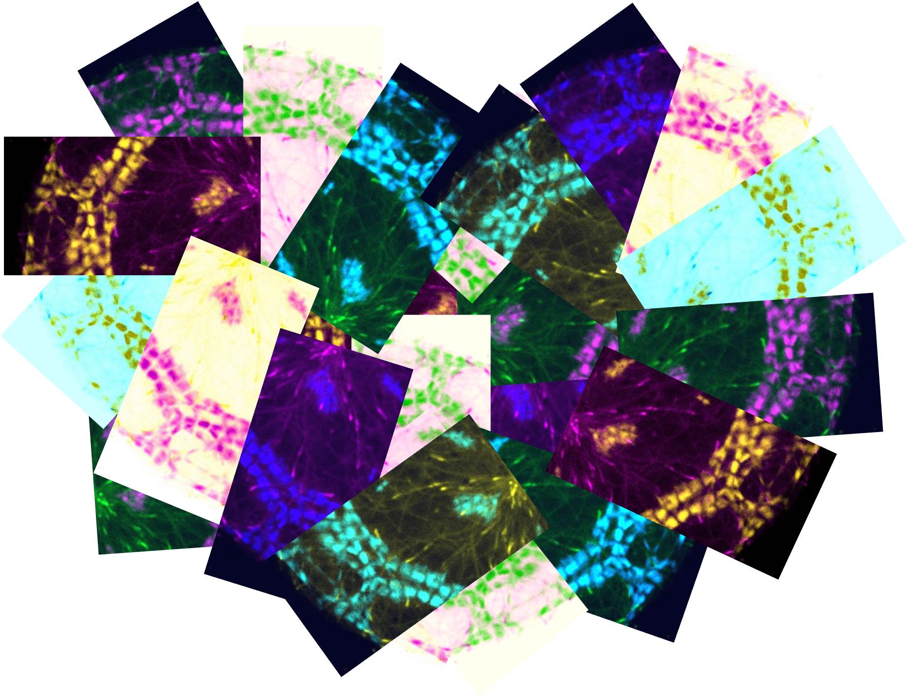
LM12
My heart beats for science
microscope: Spinning Disc 1 Nikon-Roper ILAS PhotoAblation (Kruyt building)
description: This is a compilation of pictures to form a heart shape. The pictures show a zoom of a human induced-pluripotent stem cell-derived cardiomyocyte, which was transduced with lentiviral vectors to express a GFP-tagged microtubule plus-end marker (end-binding protein 3 (EB3)) and tagRFP-LifeAct; an actin marker. In cardiac cells the actin cytoskeleton forms contractile units named sarcomeres, which allow the cell to beat
Author: Babet van der Vaart
LM14
Sun is a droplet
microscope: Nikon upright Ni microscope
description:
Author: Chao Yang
Electron microscopy

EM01
Air waves
microscope: JEOL 1011 electron microscope
description: EM image of a part of a tracheole, a branch of the insect airway system, between the circular and longitudinal muscle cell layers of the Drosophila intestinal wall
Author: Ann De Mazière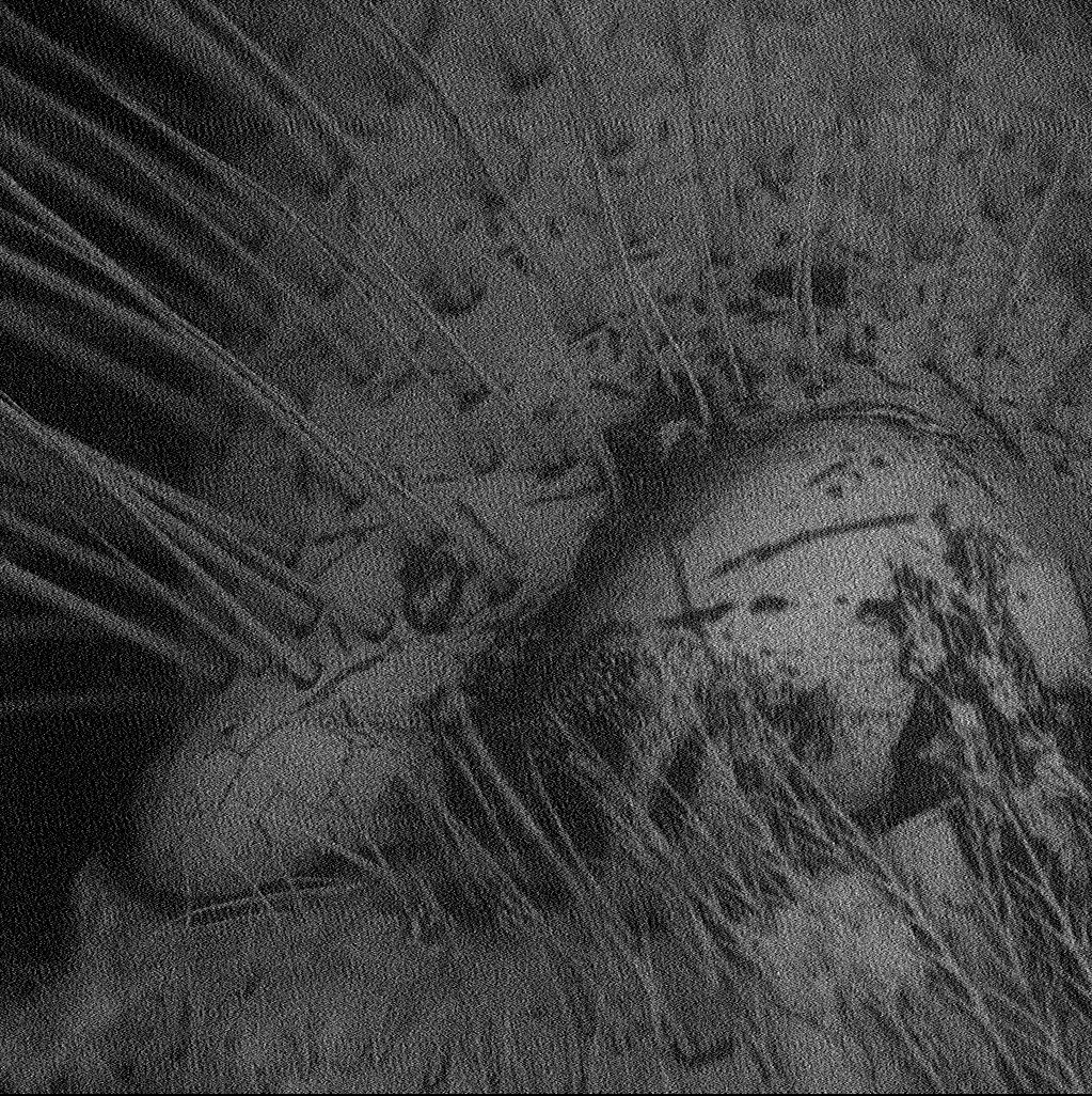
EM02
The beginning and the end
microscope: Scanning EM
description: The picture came from observing the head of a bee, more specifically the eye
Author: Ines Martins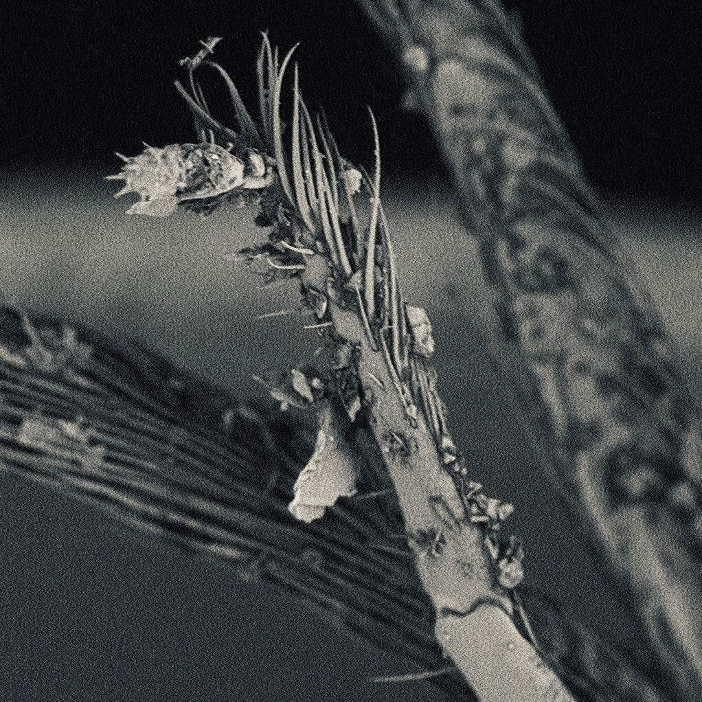
EM03
Magic Forest
microscope: Phenom PRO desktop scanning electron microscope
description: It is a 360x magnification of antennas of a bee
Author: Xudong Ouyang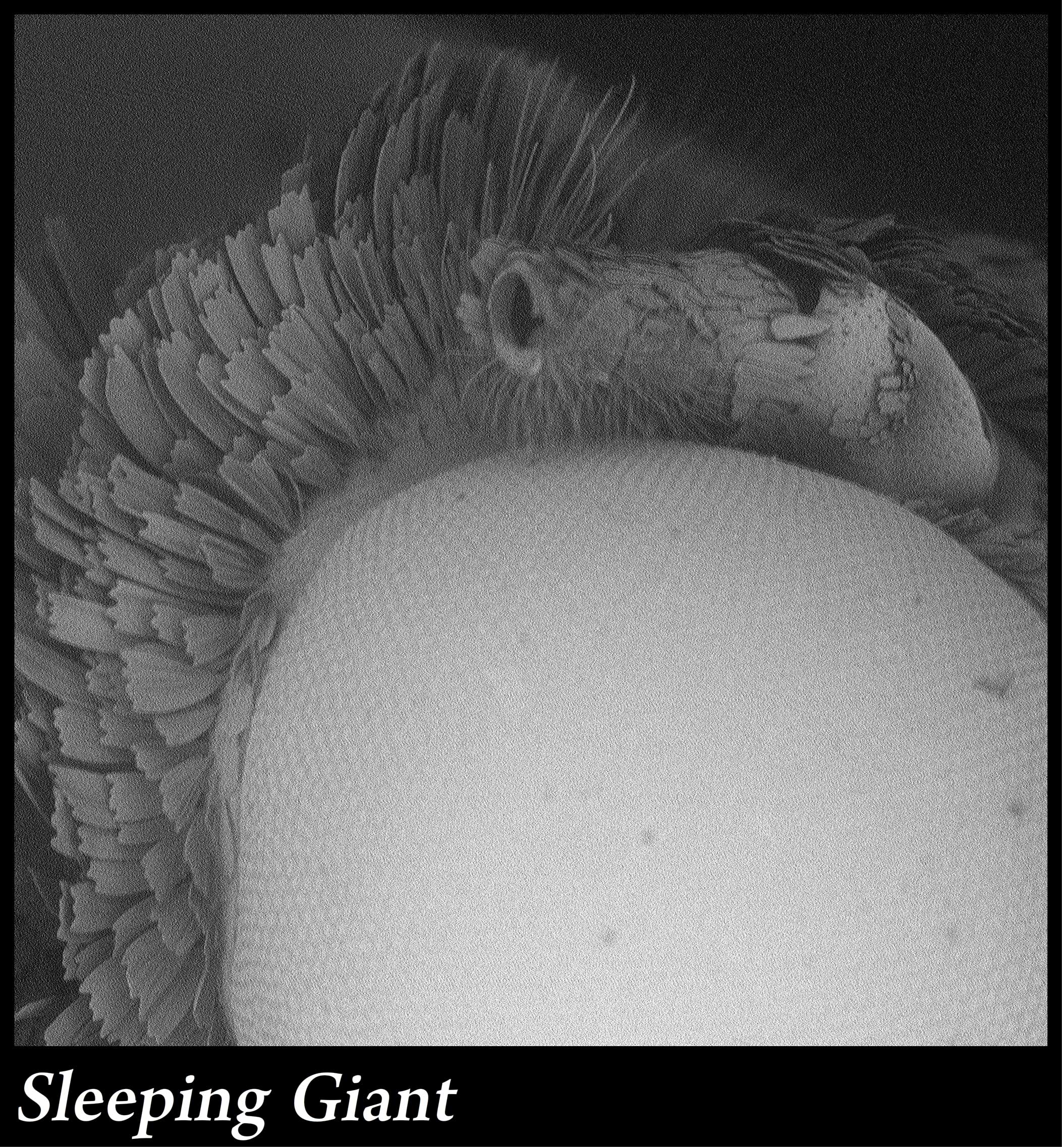
EM04
Sleeping Giant
microscope: Phenom Desktop SEM
description: 280x magnification of the eye of a moth
Author: Paul Stege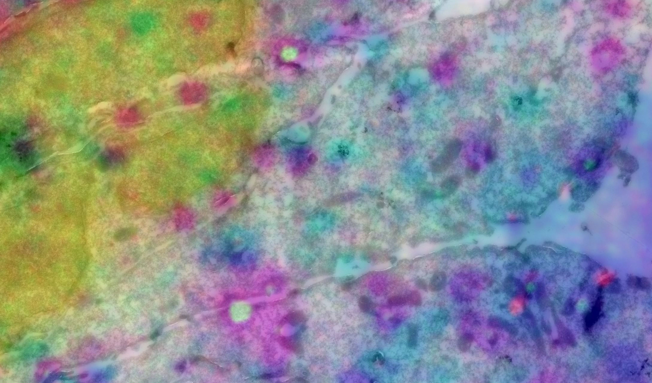
EM05
Cells and staining in Las Vegas
microscope: Tecnai 12 / DeltaVision widefield microscope
description: It occurred in some photoshop experimentation to find the best way of overlaying Fluorescence and EM images, it’s an inverted color set. While not very informative, it makes a very colorful image. It reminded me of the many colorful lights in Las Vegas. The EM picture is a stitch made with serial EM software on a Tecnai 12, the fluorescence is from a DeltaVision widefield microscope. I’d like to place it in the category ‘Electron microscopy’ images of the competition
Author: Jan van der Beek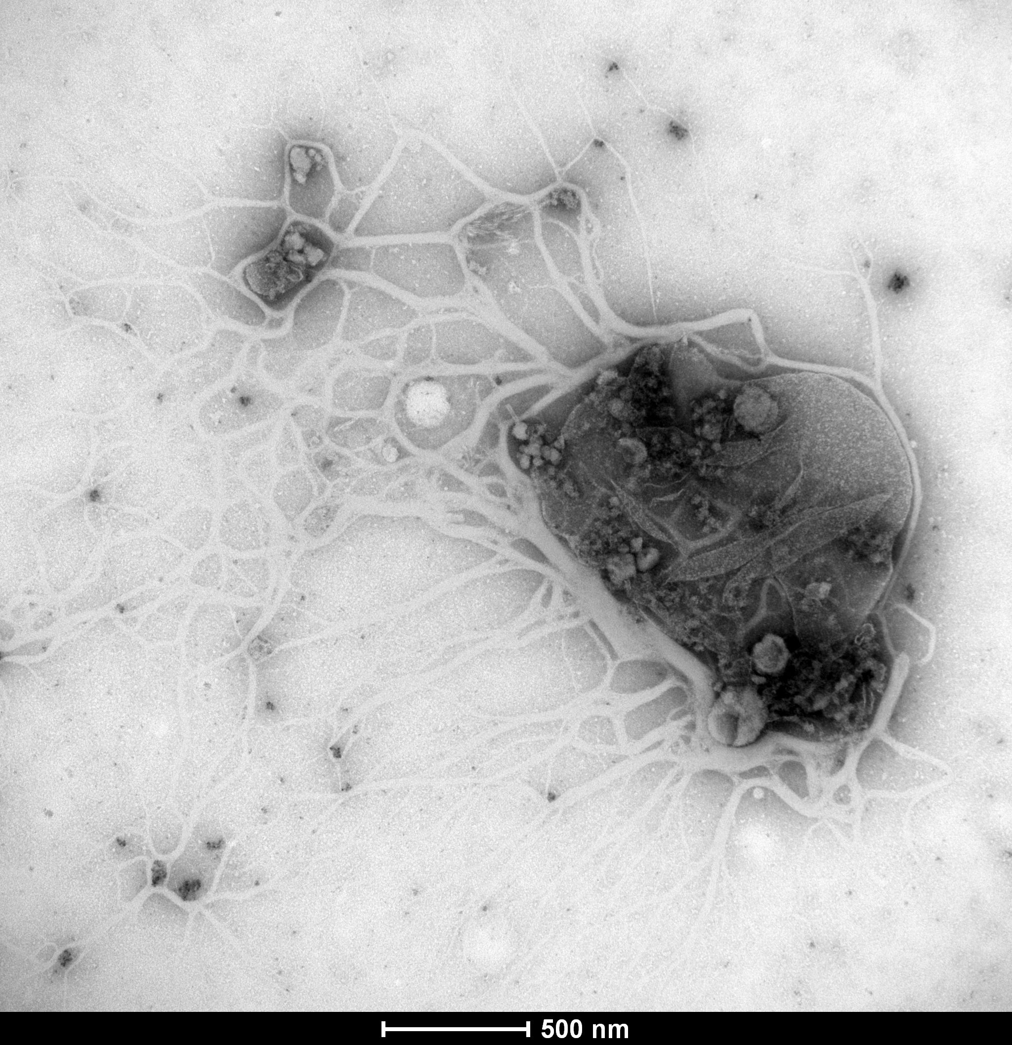
EM06
The excessive cruelty of antibiotics on bacteria
microscope: Tecnai T12
description: A new antibiotic compound appears to rip apart the bacterial cell wall and leave only the mutilated bacterial remains behind… No survivors were found in this sample
Author: Marco Viveen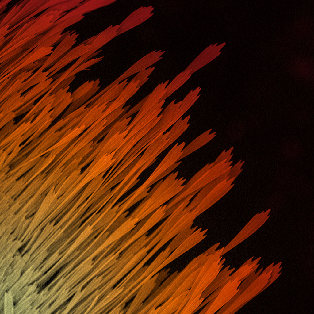
EM07
Rising Phoenix
microscope: Phenom PRO desktop scanning electron microscope
description: A 320x magnification of the wing of a moth
Author: Samantha van der Beek