Confocal Laser Scanning Microscope Carl Zeiss LSM 700
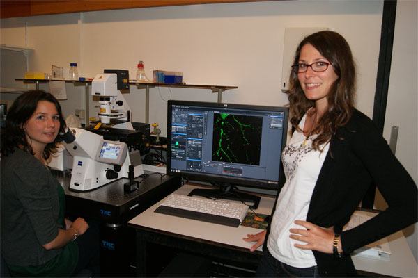
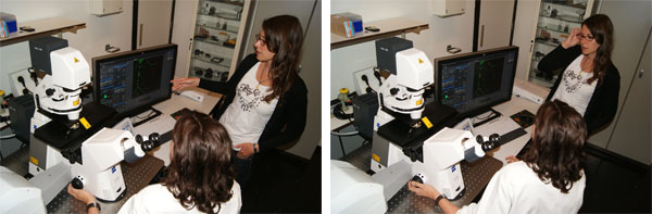
Marijn Kuijpers and Laura Gumy are studying microtubules in the growth cone during axon regeneration
Primary tasks:
- Confocal microscopy
- 3D imaging
- FRAP
Microscope: AxioObserver Z1
Objectives:
Plan-Apochromat 20x/0.8 (WD=0.55mm)
EC Plan-Neofluar 40x/1.30 Oil DIC (WD=0.21mm)
Plan-Apochromat 63x/1.40 Oil DIC (WD=0.19mm)
Laserlines: 405nm, 488nm, 555nm en 633 nm
Software: ZEN 2011
Contact:
Dr. Ilya Grigoriev
The head of the Biology Imaging Center
Division of Cell Biology, Department of Biology, Faculty of Science, Utrecht University
Room O-504
Kruytgebouw,
Padualaan 8, 3584 CH Utrecht,
The Netherlands
+31-30-2533297
I.S.Grigoriev@uu.nl
http://cellbiology.science.uu.nl/biology-imaging-center-bic