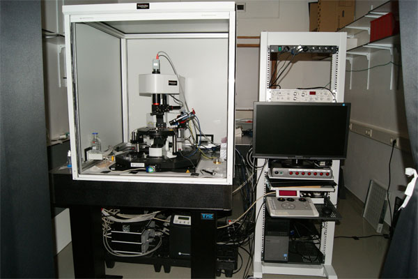
Two-Photon microscope Femtonics
Primary tasks:
Two-photon microscopy, calcium imaging
in combination with single-cell electroporation and/or electrophysiology
Microscope:
Femtonics 2D upright microscope (based on Olympus BX61 WI microscope)
Objectives:
Nikon CFI Apochromat 60X NIR water immersion
Laser:
SpectraphysicsMaiTai HP ultrafast femtosecond laser
Tuning range 690-1040 nm, pulse width <100 fs, 80 MHz repetition rate
Scanner:
Fast galvanoscanners with digital servo drivers
Detectors:
2 sub-stage (transmitted) and 2 upper (reflected) PMTs for high sensitivity
HamamatsuGaAs photomultiplier tubes (PMTs)
1 transmitted IR detector for recognizing of unstained objects (e.g. cell bodies, pipette, stimulator)
Beamsplitter(in front of PMTs):
Green PMT detection band: 475-575 nm
Red PMT detection band: 600-675 nm
Stage:
Luigs&Neumann 380FM-2P (computer-controlled) + keypad SM-5
Optical Table:
TMC
Software:
Femtonics modular measurement control and analysis software “MES” (Matlab-based)
Other equipment:
Luigs&Neumann micromanipulator UNIT Junior LE with controller SM7 for 5 axes
Axon Axopatch 200B patch clamp amplifier
Axoporator 800A single-cell electroporator
Narishige pipette puller PC-10
Contact:
Dr. Ilya Grigoriev
The head of the Biology Imaging Center
Division of Cell Biology, Department of Biology, Faculty of Science, Utrecht University
Room O-504
Kruytgebouw,
Padualaan 8, 3584 CH Utrecht,
The Netherlands
+31-30-2533297
I.S.Grigoriev@uu.nl
http://cellbiology.science.uu.nl/biology-imaging-center-bic