EM01
Air waves
microscope: JEOL 1011 electron microscope
description: EM image of a part of a tracheole, a branch of the insect airway system, between the circular and longitudinal muscle cell layers of the Drosophila intestinal wall
Author: Ann De Mazière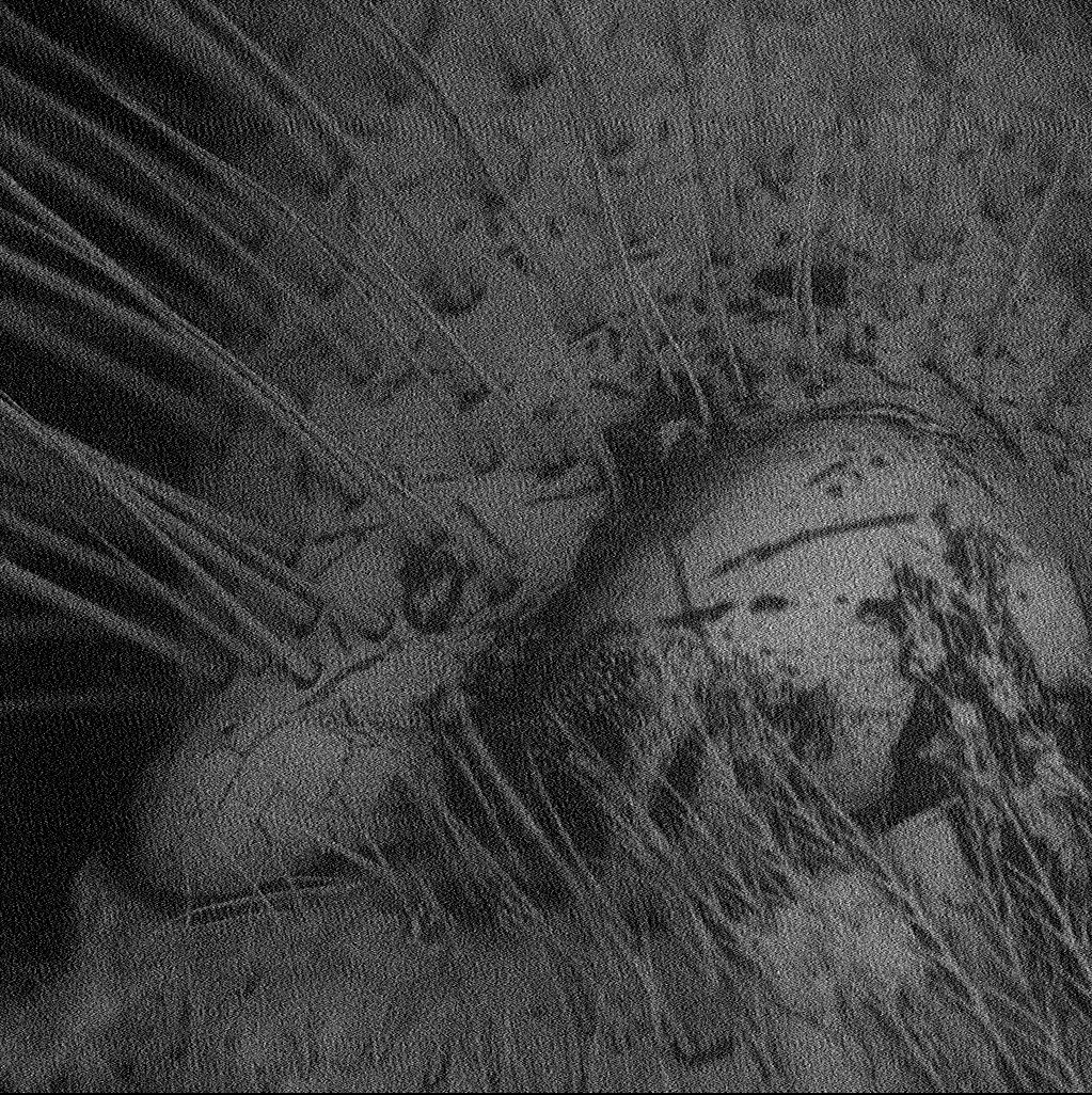
EM02
The beginning and the end
microscope: Scanning EM
description: The picture came from observing the head of a bee, more specifically the eye
Author: Ines Martins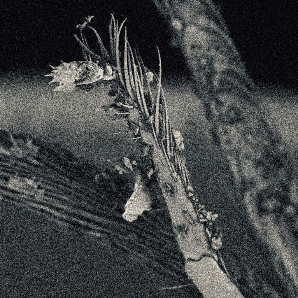
EM03
Magic Forest
microscope: Phenom PRO desktop scanning electron microscope
description: It is a 360x magnification of antennas of a bee
Author: Xudong Ouyang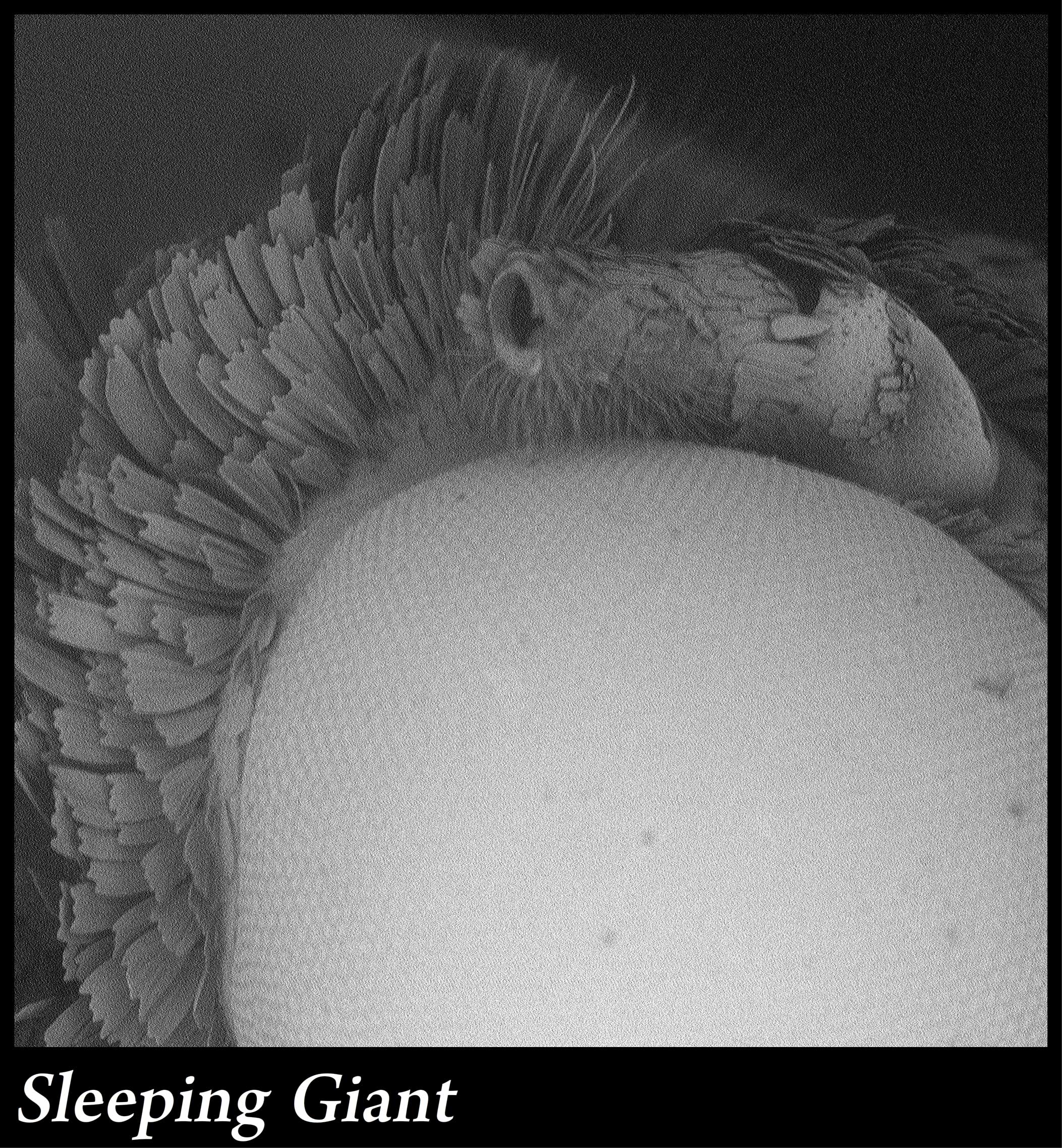
EM04
Sleeping Giant
microscope: Phenom Desktop SEM
description: 280x magnification of the eye of a moth
Author: Paul Stege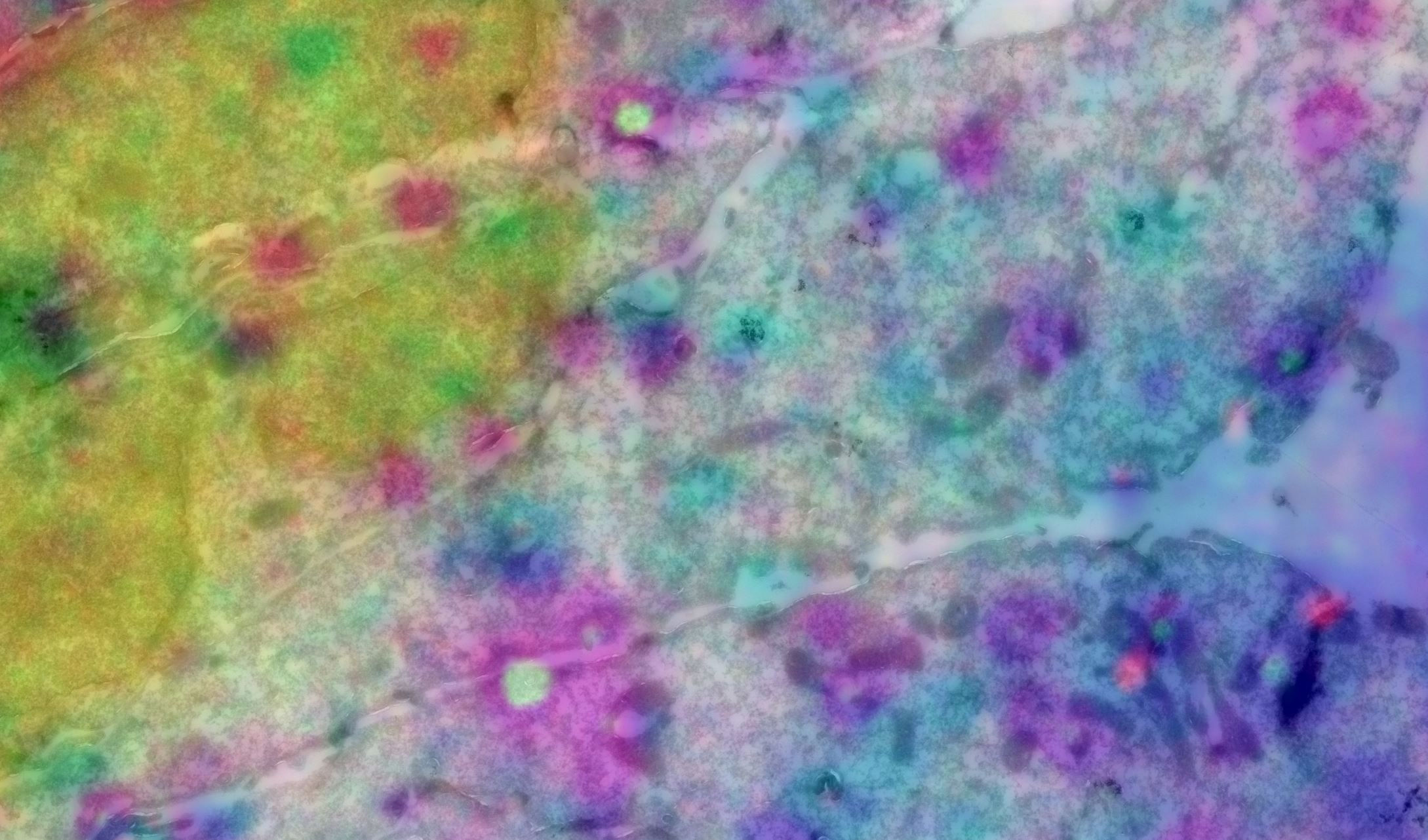
EM05
Cells and staining in Las Vegas
microscope: Tecnai 12 / DeltaVision widefield microscope
description: It occurred in some photoshop experimentation to find the best way of overlaying Fluorescence and EM images, it’s an inverted color set. While not very informative, it makes a very colorful image. It reminded me of the many colorful lights in Las Vegas. The EM picture is a stitch made with serial EM software on a Tecnai 12, the fluorescence is from a DeltaVision widefield microscope. I’d like to place it in the category ‘Electron microscopy’ images of the competition
Author: Jan van der Beek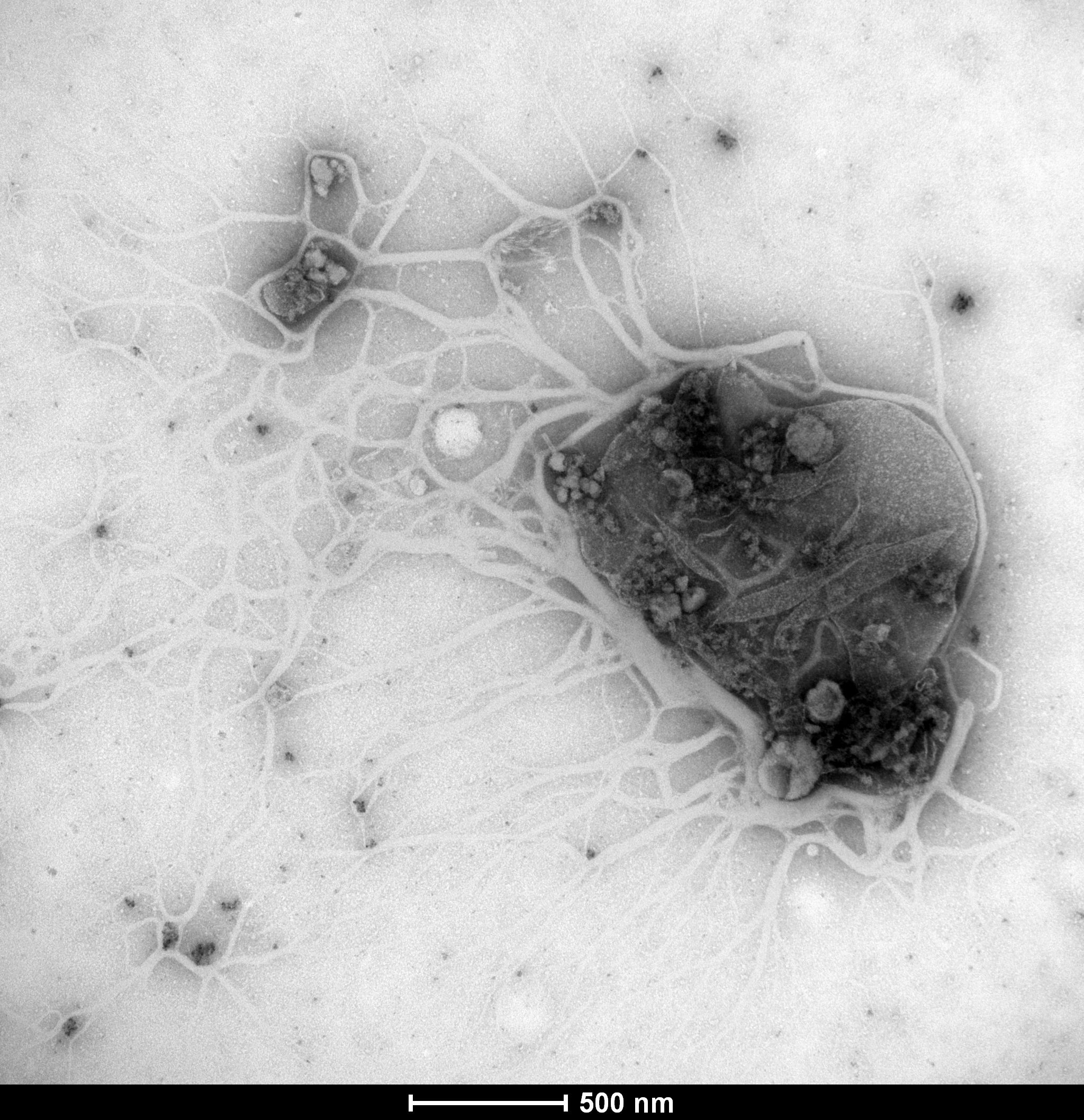
EM06
The excessive cruelty of antibiotics on bacteria
microscope: Tecnai T12
description: A new antibiotic compound appears to rip apart the bacterial cell wall and leave only the mutilated bacterial remains behind… No survivors were found in this sample
Author: Marco Viveen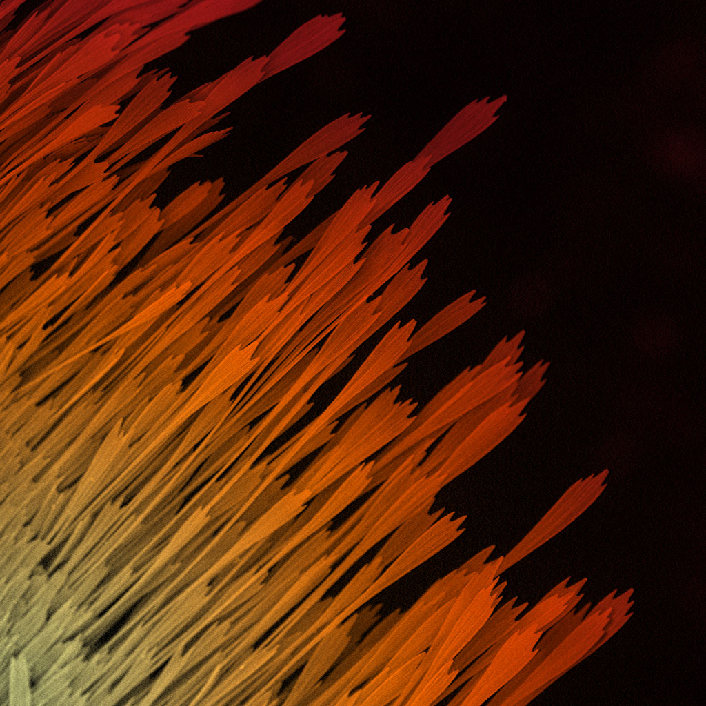
EM07
Rising Phoenix
microscope: Phenom PRO desktop scanning electron microscope
description: A 320x magnification of the wing of a moth
Author: Samantha van der Beek